Body Systems
Anatomy provides the structural framework required for physiology, pathology, surgery, radiology, diagnosis, and clinical medicine.
Advanced Anatomical Science
Advanced anatomical analysis using cutting-edge imaging and AI-driven diagnostics, transforming medical education, clinical diagnostics, surgical planning, and precision healthcare.
Abstract
Human anatomy studies the structure, organization, and relationships of the body's systems, tissues, and organs. Modern anatomy now blends cadaveric knowledge with imaging, computational modeling, AI, and immersive simulation.
Anatomy provides the structural framework required for physiology, pathology, surgery, radiology, diagnosis, and clinical medicine.
MRI, CT, ultrasound, PET, X-ray, and 3D reconstruction reveal internal anatomy without invasive exploration.
AI-driven image analysis supports segmentation, disease detection, anatomical mapping, predictive modeling, and precision diagnostics.
Parts I-II
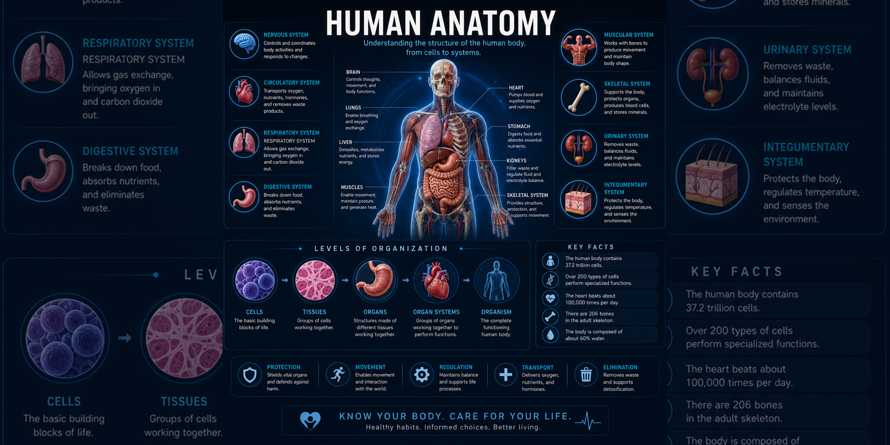
The body is organized hierarchically from cells to tissues, organs, organ systems, and the whole organism.
Large-scale structures visible to the naked eye, including organs, muscles, bones, vessels, and nerves.
Histology and cellular organization reveal tissues and microstructures that support organ function.
Applies structural knowledge to diagnosis, surgery, imaging interpretation, procedural planning, and patient care.
Covers body surfaces and lines internal organs.
Provides support and includes bone, cartilage, blood, adipose tissue, and structural matrices.
Generates movement, posture, pressure, and force through skeletal, smooth, and cardiac muscle.
Specialized for signal transmission, sensory processing, coordination, and communication.
Part III
Non-invasive imaging made it possible to visualize internal anatomy with extraordinary precision.
Foundational radiography remains essential for skeletal anatomy, fracture evaluation, thoracic conditions, and dental imaging.
CT produces high-resolution cross-sectional images that reveal bone, trauma, vascular structures, and complex anatomy.
MRI provides excellent soft-tissue contrast for brain, spine, joints, organs, tumors, and neuroanatomical mapping.
Real-time imaging supports obstetrics, vascular evaluation, cardiac assessment, procedural guidance, and soft-tissue analysis.
Metabolic imaging reveals functional activity, cancer spread, neurologic disorders, and physiologic changes beyond structure alone.
Quantitative image features can be extracted and analyzed to identify patterns linked to diagnosis, prognosis, and treatment response.
Part IV
Computational imaging creates detailed three-dimensional models that improve education, simulation, implant design, and surgical preparation.
Part V
Machine learning and deep learning algorithms can analyze imaging data, identify subtle patterns, map structures, and support diagnostic decisions.
AI outlines anatomical structures with improved speed, accuracy, and reproducibility.
Deep learning systems detect abnormalities that may be difficult for human observers to identify.
Large imaging datasets can support forecasts of disease progression, surgical outcomes, and anatomical changes over time.
Parts VI-VIII
Advanced anatomical analysis has transformed clinical practice across neuroscience, cardiovascular medicine, orthopedics, oncology, education, simulation, and surgical planning.
Advanced neuroimaging allows detailed mapping of brain anatomy, connectivity, and function.
Cardiac and vascular imaging support diagnosis, risk assessment, intervention planning, and monitoring.
Structural analysis of bone, muscle, cartilage, tendons, ligaments, and joints informs diagnosis and rehabilitation.
Digital platforms allow anatomy to be taught through immersive, interactive, patient-specific experiences.
AI-driven anatomical diagnostics require strong governance, transparency, and equitable access.
Part IX
The future of anatomical science integrates imaging, AI, robotics, genomics, and precision medicine.
Dynamic computational models simulate anatomy, physiology, and disease progression for personalized treatment planning.
Personalized anatomical models can tailor surgical, diagnostic, and therapeutic decisions to each patient's structure.
AI may provide continuous anatomical analysis during surgery and procedures to enhance precision and reduce error.
AR-guided surgery can overlay anatomical information directly onto patients for navigation and precision.
Mathematics, imaging science, and AI can identify anatomical variation and disease-related structural change across populations.
References
Akkus, Z., et al. (2017). Deep Learning for Brain MRI Segmentation: State of the Art and Future Directions. Journal of Digital Imaging, 30(4), 449-459.
Esteva, A., et al. (2019). A Guide to Deep Learning in Healthcare. Nature Medicine, 25(1), 24-29.
Mayo Clinic. (2025). PET Scan Overview.
National Institutes of Health. (2025). Magnetic Resonance Imaging.
National Library of Medicine. (2025). The Visible Human Project.
Radiological Society of North America. (2025). Computed Tomography Scans and Safety.
Rizzo, S., et al. (2018). Radiomics: The Facts and the Challenges of Image Analysis. European Radiology Experimental, 2(1), 36.
Sutherland, J., et al. (2021). Virtual Reality and Anatomy Education: A Systematic Review. Anatomical Sciences Education, 14(5), 700-709.
Topol, E. (2019). High-Performance Medicine: The Convergence of Human and Artificial Intelligence. Nature Medicine, 25(1), 44-56.
World Health Organization. (2024). Ethics and Governance of Artificial Intelligence for Health.
FAQ
Evidence-based answers to common questions about anatomical organization, imaging, 3D anatomy, AI analysis, and clinical anatomical variation.
The body is organized from atoms and molecules to organelles, cells, tissues, organs, organ systems, and the whole organism. The major systems include integumentary, skeletal, muscular, nervous, endocrine, cardiovascular, lymphatic, respiratory, digestive, urinary, and reproductive systems.
Modern anatomy uses X-ray, CT, MRI, ultrasound, PET, angiography, 3D reconstruction, radiomics, and digital visualization platforms to study internal structures non-invasively.
3D digital anatomy reconstructs imaging data into interactive anatomical models for education, simulation, surgical planning, implant design, and patient-specific care.
AI can segment tissues, detect disease patterns, measure organ volumes, map vasculature, predict anatomical changes, and support clinical decision-making from imaging data.
Anatomical variations can affect diagnosis, surgery, procedural safety, imaging interpretation, device placement, and personalized treatment planning.